Airway blocks: superior laryngeal and transtracheal (2027)
Stem
At the ultrasound station, the scenario card describes a 58-year-old man, 78 kg, with a floor-of-mouth tumor and limited mouth opening, scheduled for awake flexible-scope intubation. The standardized patient lies supine with a pillow under his shoulders. The examiner at the machine reads the nerve block task: the internal branch of the superior laryngeal nerve on the right. Linear and curvilinear probes are available.
Question
Show me where you block the superior laryngeal nerve: where is your probe, which structures do you name, and where does your needle tip go?
Ideal answer
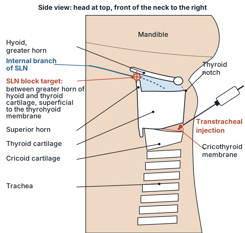
HeadlineMy target is the space where the internal branch pierces the thyrohyoid membrane, between hyoid and thyroid cartilage; the tip goes just superficial to it.1
- High-frequency linear probe, shallow depth, because the targets are superficial.
- Midline sagittal on the thyroid cartilage, then slide right. Neck slightly extended;2 I settle parasagittal just below the angle of the mandible, marker toward his head.
- Name the two bright, shadowing structures. Greater horn of the hyoid cephalad, thyroid cartilage caudad; between them the thyrohyoid muscle superficial, the membrane deep, pre-epiglottic fat behind.1
- Show the tip: under the muscle, above the membrane. Out of plane, 1–2 mL of lidocaine, then the same on the left;1,2 the nerve is usually too small to see, so the space is my target.1
- Then I stop and ask for the freeze, because I have two and a half minutes.3
Breaking down the answer
- It is a 2027 target — the outline adds superior laryngeal and transtracheal blocks, and airway structures from the hyoid to the trachea, from 2027; its new airway management section lists landmarks for airway blocks.4 Usual template: probe orientation, two structures, needle-tip location.3
- One target per task — extra anatomy costs time against the 2 min 30 s limit.3 Changes if: the image is crowded, when I go back to the midline thyroid cartilage and slide out again.2
- Aspirated air means too deep — the tip is through the membrane into the airway, so I withdraw before injecting; a tip deep to the membrane without air may sit in the pre-epiglottic space.
- Know what it covers — the internal branch numbs the larynx above the cords, the epiglottis and the base of the tongue;1 below the cords comes from the recurrent laryngeal nerve, which the transtracheal injection reaches.2
- Count the lidocaine — all routes add up, with a ceiling of 9 mg/kg of lean body weight; superior laryngeal blocks have been associated with higher plasma lidocaine levels and with toxicity.5
| Block | Probe and view | Two structures | Needle tip |
|---|---|---|---|
| Superior laryngeal (internal branch) | Linear, parasagittal, below the mandibular angle | Greater horn of hyoid; thyroid cartilage | Deep to thyrohyoid muscle, superficial to thyrohyoid membrane1 |
| Transtracheal | Linear, midline sagittal | Thyroid cartilage; cricoid cartilage | Through the cricothyroid membrane into the airway2 |
Follow-up question
The examiner moves to the next scenario: the same patient now needs a transtracheal injection, and his neck is thick enough that you cannot palpate the cricothyroid membrane. Where does your needle go, and how do you show it?
Follow-up answer
HeadlineThe needle goes through the cricothyroid membrane in the midline; I find it with the linear probe longitudinal over the trachea.2
I slide toward his head until the tracheal rings give way to the cricoid, a larger hypoechoic oval, with the thyroid cartilage cephalad. The cricothyroid membrane is the bright line joining them, with the air-mucosa line deep to it. I name both cartilages, center the membrane and mark the skin. The tip goes through it in the midline, aspirating air to confirm the trachea, with 2–3 mL of 2% lidocaine on expiration; the cough spreads it to the cords and trachea.2,1 If he is very obese, I switch to a curvilinear probe for depth.
Pitfalls that cost points
- Calling the cricoid the thyroid cartilage because the scan started too low.
- Forgetting to add the transtracheal, nebulized and topical lidocaine to the block dose.
Rapid-fire
Q: Which nerve does the superior laryngeal block target, and from where? A: The internal branch, a vagal branch that passes just below the greater horn of the hyoid toward the thyrohyoid membrane.1
Q: What surrogate marker helps in a transverse view? A: The superior laryngeal artery, which runs with the internal branch.1
Q: How do tracheal rings look in a longitudinal view? A: A row of hypoechoic rounded cartilages in front of a bright air line, often called a string of beads.
Q: Why do DAS guidelines reserve invasive blocks? A: They raise plasma lidocaine, have caused toxicity and can be less comfortable than topical techniques.5
Q: Which other airway nerve block is not on the outline? A: The glossopharyngeal block; the outline names only superior laryngeal and transtracheal.4
Numbers to know
| Item | Value |
|---|---|
| Testing starts | 20274 |
| Time per task | ≤2 min 30 s3 |
| Probe | Linear, 6–13 MHz2 |
| SLN dose per side | 1 mL of 2% or 2 mL of 1.5% lidocaine1,2 |
| Transtracheal | 2–3 mL of 2% lidocaine2,1 |
| Lidocaine ceiling | 9 mg/kg lean body weight5 |
Links
- The ultrasound application station: format and tasks, Awake fiberoptic intubation: topicalization and sedation, Airway station: exam, algorithm, device choice (2027), Cannot intubate, cannot oxygenate, Local anesthetic systemic toxicity
- ABA OSCE Content Outline (airway blocks and airway structures from 2027); DAS guidelines for awake tracheal intubation in adults (2020)
Sources
- 1.Comparison of ultrasound and anatomical landmark-guided technique for superior laryngeal nerve block to aid awake fibre-optic intubation: a prospective randomised clinical study · Indian Journal of Anaesthesia, 2017 · ebi.ac.ukAmbi US et al. Parasagittal submandibular probe; SLN space bounded by hyoid, thyroid cartilage, thyrohyoid muscle and thyrohyoid membrane; 1 mL 2% lidocaine per side out of plane; transtracheal 3 mL 2%.
- 2.Comparison of landmark-guided and ultrasound-guided technique for superior laryngeal nerve block to aid fibreoptic intubation: an observational study · Journal of Anaesthesiology Clinical Pharmacology, 2023 · ebi.ac.ukMagadum N, Nileshwar A et al. Linear 6-13 MHz probe, midline sagittal then lateral; 2 mL 1.5% lidocaine per side; cricothyroid membrane found by ultrasound, transtracheal 2 mL 2%.
- 3.OSCE Example Scenarios (all stations) · ABA, 2023 · theaba.orgUltrasound station: examiner runs the machine, 3 tasks, no more than 2 min 30 s each; nerve block task = probe orientation, two structures, needle-tip location.
- 4.APPLIED Exam Objective Structured Clinical Examination (OSCE) Content Outline · ABA · theaba.orgNerve block list adds 'Airway (superior laryngeal, transtracheal)' with testing to start 2027; airway-related structures (trachea, tracheal rings, cricoid, cricothyroid membrane, thyroid cartilage, vocal cords, hyoid, epiglottis, tongue, esophagus) also from 2027.
- 5.Difficult Airway Society guidelines for awake tracheal intubation (ATI) in adults · DAS, 2020 · ebi.ac.ukFull text via Europe PMC (PMC7078877). Topical lidocaine maximum 9 mg/kg lean body weight; SLN and glossopharyngeal blocks linked to higher plasma levels and LAST.
Facts on this page were checked against these sources on Oct 16, 2026. Guidelines change; confirm doses against current institutional protocols.